Back Muscles Anatomy Ct : The 6 Best Muscles To Self Massage For Instant Relief Of Neck And Upper Back Tension Head To Toe Muscle Clinic : This study included 31 patients, mean age 47 years, with chronic low back pain.
byAdmin•
0
Back Muscles Anatomy Ct : The 6 Best Muscles To Self Massage For Instant Relief Of Neck And Upper Back Tension Head To Toe Muscle Clinic : This study included 31 patients, mean age 47 years, with chronic low back pain.. Use the mouse scroll wheel to move the images up and down alternatively use the tiny arrows (>>) on both side of the image to move the images.>>) on both side of the image to move the images. On anatomical parts the user can choose to display the various structures in colored illustrations of the anatomy of the back and spine: The rhomboid muscle is activated as you bring and squeeze your scapula or shoulder blades back and together. This group is made of three subgroups, with the group divisions occurring by location. The calf muscle, on the back of the lower leg, is actually made up of two muscles:
Interobserver reliability was not investigated in the current study. Posted by radiologypics ⋅ march 21, 2013 ⋅ 1 comment. Anatomy of the lumbar spine (ct scan) this anatomy module is dedicated to interns and students that wish to learn more about the anatomy of the lumbar spine in ct. The gastrocnemius is the larger calf muscle, forming the bulge visible beneath the skin. The back muscles are anatomically layered into superficial (extrinsic) and deep (intrinsic) muscles.
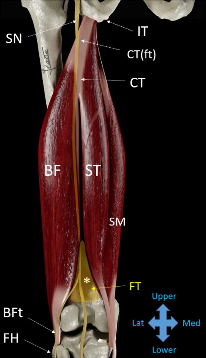
Sonographic Landmarks In Hamstring Muscles Springerlink from media.springernature.com The back muscles are anatomically layered into superficial (extrinsic) and deep (intrinsic) muscles. Superficial back muscles, intermediate back muscles and intrinsic back muscles.the intrinsic muscles are named as such because their embryological development begins in the back, oppose to the superficial and intermediate back muscles which develop elsewhere and are therefore classed as extrinsic muscles. Each block is separated by a disc that sits in between and each vertebra has a facet joint on either side. Along with sternocleidomastoid muscle, it is invested by the superficial layer of the deep cervical fascia, which splits around it. On anatomical parts the user can choose to display the various structures in colored illustrations of the anatomy of the back and spine: The rhomboid muscle is activated as you bring and squeeze your scapula or shoulder blades back and together. The erector spinae group is the intermediate layer of the intrinsic muscles of the back. The trapezius muscle (or simply trapezius, latin:
Thigh muscles are responsible for allowing normal gait and proper lower extremity function (1). The images are available in the three planes, axial, sagittal. The muscles of the back are a group of strong, paired muscles that lie on the posterior aspect of the trunk. This blog post article is an overview of the muscles of the pelvis. The trapezius muscle is a large, broad superficial muscle of the posterior neck and back. Each block is separated by a disc that sits in between and each vertebra has a facet joint on either side. They provide movements of the spine , stability to the trunk, as well as the coordination between the movements of the limbs and trunk. Musculus trapezius) is a significant muscle of the back lying in the most superficial layer.it extends from the occipital bone down to the lower thoracic vertebrae and laterally to the scapula.the trapezius moves and stabilizes the scapula. Using this atlas of human anatomy of the spine and back. The intrinsic back muscles are found deeper to the extrinsic muscles, separated from them by the thoracolumbar fascia. The back muscles are anatomically layered into superficial (extrinsic) and deep (intrinsic) muscles. Filed under anatomy, ct, head and neck. Posted by radiologypics ⋅ march 21, 2013 ⋅ 1 comment.
The extrinsic back muscles are located in the back, but act to produce movements of the shoulder and assist respiration. Related posts of muscles shoulder and back muscle anatomy lower extremity. Weak adductor muscles may cause knee instability and adductor strain (2). The intrinsic back muscles are found deeper to the extrinsic muscles, separated from them by the thoracolumbar fascia. This muscle is located on the upper portion of the back anatomy, underneath the trapezius.
Ct And Mri Determination Of Intermuscular Space Within Lumbar Paraspinal Muscles At Different Intervertebral Disc Levels from journals.plos.org This article gives an overview of the back's structure and its major muscles. The gastrocnemius is the larger calf muscle, forming the bulge visible beneath the skin. Tutorials on the anatomy and actions of the back muscles, using interactive animations, diagrams, and illustrations. Similar to learning the muscles of the lumbar spine/trunk, it can be helpful to first look at the. This study included 31 patients, mean age 47 years, with chronic low back pain. Filed under anatomy, ct, head and neck. There is a dissection assistance pdf file that you can use to assist you in your lab preparation. They start at the top of the neck and go down to the tailbone.
Vertebrae, bones, joints, ligaments, muscles, muscular system, fascia, arteries, veins, nerves and various adjacent organs.
Muscles make up a large part of the anatomy (structure) of the back. The muscles of the back are a group of strong, paired muscles that lie on the posterior aspect of the trunk. To perform clinical clinical orthopedic manual therapy to the lumbar spine. The muscles of the back. This blog post article is an overview of the muscles of the pelvis. The rhomboid muscle is activated as you bring and squeeze your scapula or shoulder blades back and together. The trapezius muscle (or simply trapezius, latin: Related posts of muscles shoulder and back muscle anatomy lower extremity. Muscle anatomy lower extremity 12 photos of the muscle anatomy lower extremity anatomy lower extremity muscle quiz, lower extremity muscle anatomy ct, lower extremity muscle anatomy mri, muscle anatomy lower extremity, muscular anatomy of lower limb, human muscles, anatomy lower extremity muscle quiz, lower. Lower back pain is a pervasive symptom. There is a dissection assistance pdf file that you can use to assist you in your lab preparation. Anatomy of the upper back muscles. Thigh muscles are responsible for allowing normal gait and proper lower extremity function (1).
Thigh muscles are responsible for allowing normal gait and proper lower extremity function (1). Lower back pain is a pervasive symptom. This mri chest (thorax) axial cross sectional anatomy tool is absolutely free to use. The trapezius muscle the trapezius is one of the major muscles of the back and is responsible for moving, rotating, and stabilizing the scapula (shoulder blade) and extending the head at the neck. Posted by radiologypics ⋅ march 21, 2013 ⋅ 1 comment.
Chest Anatomy Mri Chest Thorax Axial Anatomy Free Cross Sectional Anatomy from mrimaster.com They start at the top of the neck and go down to the tailbone. Anatomy of the lumbar spine (ct scan) this anatomy module is dedicated to interns and students that wish to learn more about the anatomy of the lumbar spine in ct. Lower back pain is a pervasive symptom. Iliocostalis subgroup is the most lateral longissimus subgroup is between iliocostalis and spinalis The muscles of the back. The gastrocnemius is the larger calf muscle, forming the bulge visible beneath the skin. Interobserver reliability was not investigated in the current study. On anatomical parts the user can choose to display the various structures in colored illustrations of the anatomy of the back and spine:
The seventh cervical vertebra, referred to as c7, meets the first of 12 thoracic vertebrae t1 at the base of the neck, a.
This blog post article is an overview of the muscles of the pelvis. The trapezius muscle the trapezius is one of the major muscles of the back and is responsible for moving, rotating, and stabilizing the scapula (shoulder blade) and extending the head at the neck. Anatomy of the lumbar spine (ct scan) this anatomy module is dedicated to interns and students that wish to learn more about the anatomy of the lumbar spine in ct. (2017, elsevier) should be consulted. This study included 31 patients, mean age 47 years, with chronic low back pain. The extrinsic back muscles are located in the back, but act to produce movements of the shoulder and assist respiration. The deep or intrinsic muscles of the back (fig. Vertebrae, bones, joints, ligaments, muscles, muscular system, fascia, arteries, veins, nerves and various adjacent organs. Back dissection, cranial neve 11, dissection, latisumus dorsi, lavator. The gastrocnemius is the larger calf muscle, forming the bulge visible beneath the skin. Back pain is common and might be caused by a problem with a muscle. Lower back pain is a pervasive symptom. Similar to learning the muscles of the lumbar spine/trunk, it can be helpful to first look at the.
Use the mouse scroll wheel to move the images up and down alternatively use the tiny arrows (>>) on both side of the image to move the images>>) on both side of the image to move the images back muscles anatomy. The muscles of the back can be arranged into 3 categories based on their location:

%20axial%20image%2014.jpg)